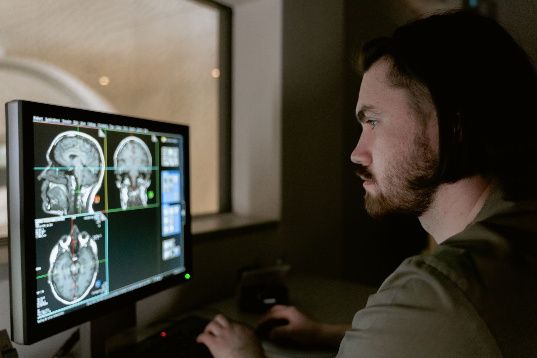
Scientists at Stanford University have reported a successful transplantation of laboratory‑grown human brain tissue into mice that were genetically engineered to lack the majority of their cerebral cortex. The findings, published in the journal Nature, describe a new animal model that could accelerate research into a range of neurological conditions.
Method and Findings
The transplanted tissue consisted of cortical organoids—three‑dimensional laboratory cultures that resemble specific regions of the brain. These organoids were created by converting human skin cells into pluripotent stem cells, which can differentiate into most cell types in the body, including neurons that form cortical circuitry. Researchers placed the organoids into specially bred lab mice whose cerebrum was largely absent, providing a biological niche for the human graft.
After implantation, the human tissue not only survived but thrived, growing and establishing functional connections with the host mouse brain and extending to the spinal cord. Images included in the study show the stark contrast between the cortical deficit in untransplanted mice and the restored brain volume once the organoid graft was in place. Senior author Professor Sergiu Pasca noted that the grafts generated a broad diversity of cortical cell types and formed working connections throughout the mouse nervous system.
Pasca emphasized that the resulting “xenocortical” mice retain a mouse nervous system while containing a larger volume of human cortical tissue that integrates and forms connections within the host. He cautioned that the animals do not reproduce the full complexity of a human brain, but they provide a platform to study human neural cell types and developmental processes that would otherwise be extremely difficult to access.
Potential Clinical Impact
The new model opens avenues for investigating disorders such as profound autism, schizophrenia, cerebral palsy and epilepsy. By observing how human cortical tissue develops in a living organism, researchers can begin to ask how disease‑associated genetic changes alter neural development and circuitry, and whether potential treatments can prevent or correct those changes.
Alison Singer, president of the Autism Science Foundation, described the work as a “critical step” toward precision medicine, noting that organoid models derived from an individual’s unique genetic makeup could reveal what goes wrong in that person’s brain. Pasca added that neuroscientists will be able to learn much more about the causes and mechanisms of neurodevelopmental and pregnancy‑incurred disorders and to test possible interventions to correct or prevent them.
Expert Commentary
Studying the living human brain has long been constrained by ethical considerations, making direct molecular and cellular investigation virtually impossible. The Stanford study demonstrates that human cortical organoids can be integrated into an in‑vivo system, offering a new experimental window while respecting ethical boundaries. The researchers anticipate that the xenocortical mice will serve as a valuable tool for dissecting the cellular underpinnings of complex brain disorders and for evaluating therapeutic strategies before they move to human trials.

Helene Elliott is the Lead Science & Space Reporter at News Raise. She reports on aerospace missions, astrophysics discoveries, quantum research, and environmental technology.