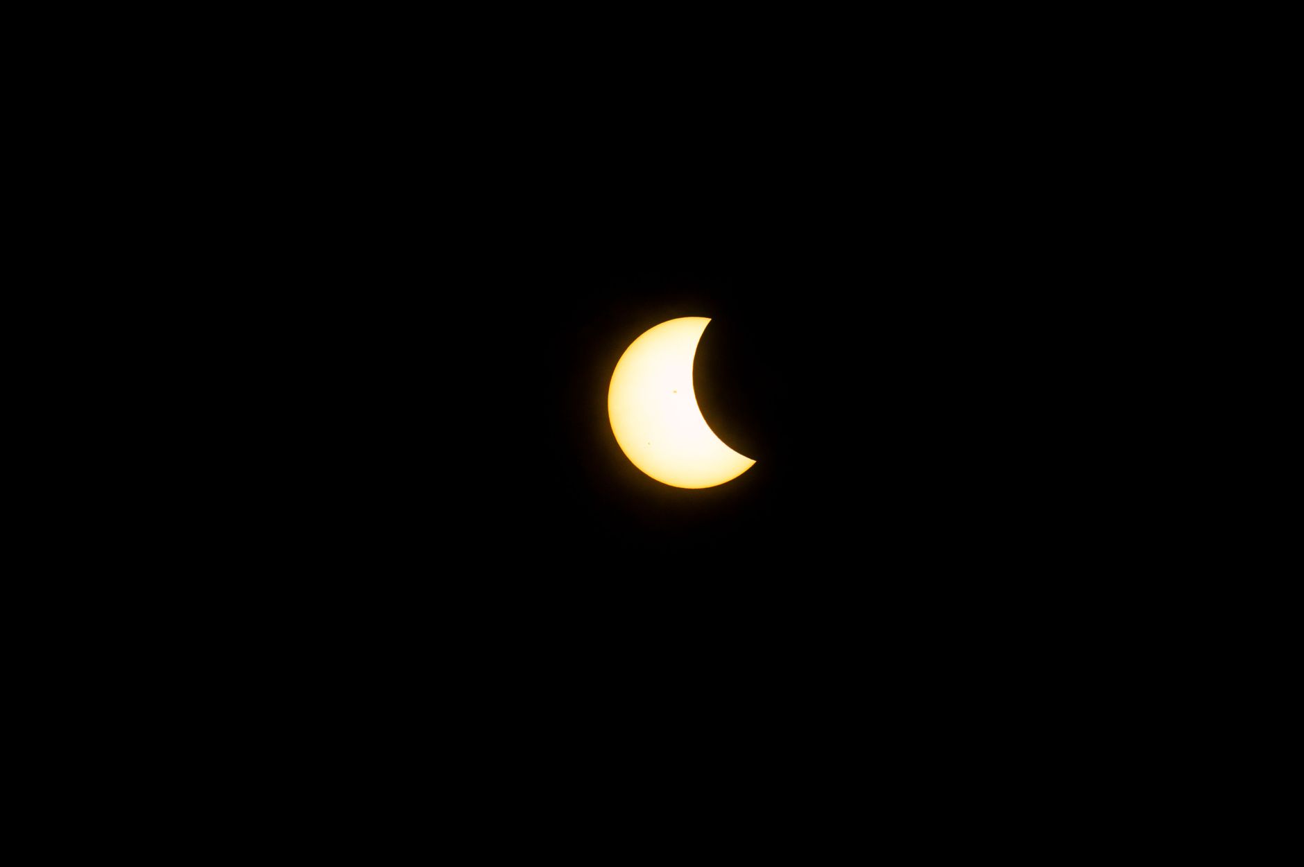
The total solar eclipse of 21 August 2017 traversed a narrow swath of the United States, but most of the country, including Staten Island, New York, experienced a partial eclipse with roughly 70 % of the Sun obscured. Tens of millions of people watched the event, many with protective glasses that met the ISO‑12312‑2 standard recommended by the American Astronomical Society.
Eyewitness exposure and immediate symptoms
Twenty‑six‑year‑old Nia Payne, a resident of Staten Island, looked at the Sun without any protection. She first glanced for about six seconds, then, feeling uneasy, borrowed a pair of glasses that appeared to be eclipse filters but later were judged by ophthalmologists to be non‑certified. She continued to stare for another fifteen to twenty seconds through the inadequate lenses. By the time she entered her home, the center of her left visual field was blurred.
Within two days the blurred spot resolved into a distinct crescent‑shaped scotoma that sat precisely where the Moon’s silhouette had been during the eclipse. The shape persisted, overlaying everything she tried to read or view.
Clinical evaluation and imaging
Payne presented to the New York Eye and Ear Infirmary of Mount Sinai several days after the eclipse. A peer‑reviewed case report published in JAMA Ophthalmology in December 2017 detailed the findings of Chris Y. Wu, Michael E. Jansen, Jorge Andrade, Toco Y.P. Chui, Anna T. Do, Richard B. Rosen and Avnish Deobhakta. Visual acuity measured 20/20 in the right eye and 20/25 in the left, a relatively mild loss by clinical standards, yet the patient reported that the crescent scotoma dominated her central vision.
To verify whether retinal damage corresponded to Payne’s drawing, clinicians employed adaptive optics scanning light ophthalmoscopy (AOSLO), a high‑resolution technique originally devised for astronomy. AOSLO corrects for corneal and lens imperfections in real time, enabling cellular‑level imaging of the retina. The scans revealed a region of destroyed photoreceptor cells in the central retina of the left eye and a milder, similarly shaped area in the right eye. Both regions were crescent‑shaped and aligned with the orientation of Payne’s hand‑drawn sketch, which mirrored the eclipse’s geometry.
Mechanism of injury and long‑term outlook
The report explained solar retinopathy as damage to photoreceptors caused by two overlapping mechanisms: thermal injury from focused sunlight raising retinal temperature beyond cellular tolerance, and photochemical injury from ultraviolet and short‑wavelength visible light breaking molecular bonds in photopigments, generating free radicals. Once photoreceptor cells are destroyed, they do not regenerate.
Dr. Avnish Deobhakta, the senior author, described the correspondence between Payne’s drawing and the adaptive optics images as “unusually direct,” noting that the eye’s own optics had projected the crescent of visible Sun onto a specific retinal region, resulting in a matching pattern of cellular loss.
Since the case report, Payne’s condition has neither significantly improved nor deteriorated. Solar retinopathy has no known treatment, and the permanent scotoma remains. She has adapted by favoring her right eye for most tasks, but reading remains difficult and she must hold screens close to compensate for the central visual defect.
The case underscores the importance of using certified eclipse glasses and illustrates how modern imaging can directly link a visual symptom to a precise retinal injury pattern.

Norman Pearlstine is the Executive Editor and Co-Founder at News Raise. With over two decades of experience across financial journalism, corporate governance, and market analysis, Norman leads the editorial direction and ensures strict adherence to journalistic accuracy and ethics.