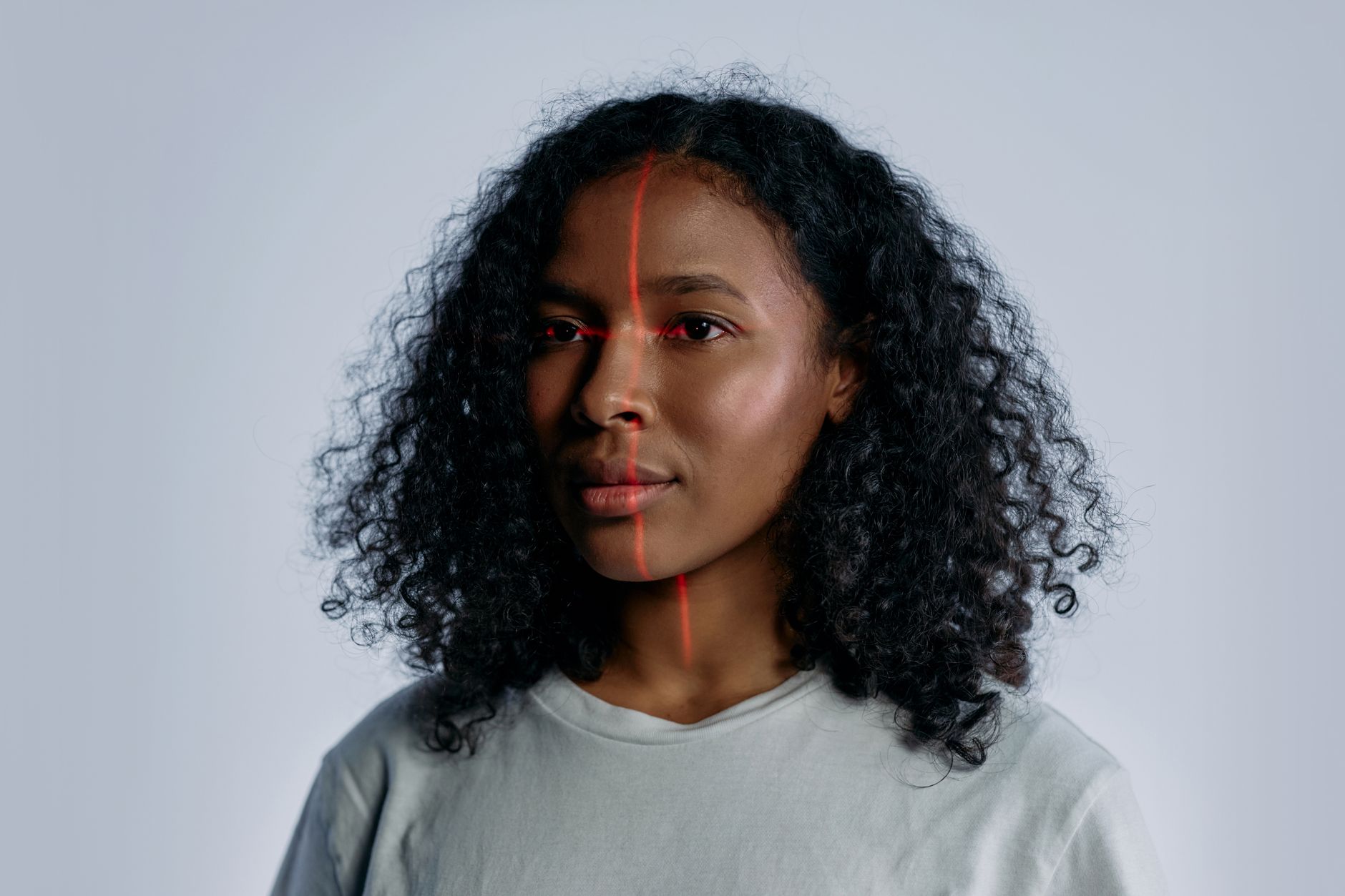
Scientists affiliated with Mass General Brigham have introduced an artificial‑intelligence platform that can infer a cancer patient’s likelihood of survival from a single photograph of the face. The approach, described in a study published in Lancet Digital Health, leverages a massive dataset of more than 40 million facial images spanning diverse ages and ethnicities to train a foundational model that is later fine‑tuned for health‑related predictions.
How the AI System Works
The technology comprises two complementary algorithms derived from the same underlying model. The first, called FAHR‑FaceAge, estimates a person’s “biological age” – the age the face appears to represent – and then relates that estimate to cancer outcomes. The second, FAHR‑FaceSurvival, bypasses the age estimate and directly predicts survival risk from the image. Although both tools share a common training base, each focuses on distinct facial regions. FAHR‑FaceAge gives weight to the nasolabial creases, the forehead just above the eye sockets, and the temples, while FAHR‑FaceSurvival concentrates on the area beneath the eyes and around the nasal bridge.
Lead co‑author Hugo Aerts highlighted that the model’s ability to learn a general facial representation from a huge image corpus enables rapid adaptation to multiple health biomarkers without rebuilding each model from scratch.
Clinical Findings and Implications
When applied to a cohort of U.S. cancer patients receiving radiotherapy, the system revealed a clear link between perceived facial age and mortality risk. Individuals whose faces appeared five or more years older than their chronological age faced a 21 % higher chance of death over an average four‑year follow‑up, whereas those who looked five or more years younger experienced a 17 % reduction in risk.
Beyond age estimation, the direct survival predictor demonstrated that facial cues alone can encode prognostic information. Aerts noted that this marks the first demonstration of an AI model directly forecasting cancer survival from a simple photograph.
Radiation oncologist Dr. Raymond Mak emphasized the potential clinical value: as the population ages, physicians need objective measures of physical reserve and frailty before recommending intensive therapies such as radiation, surgery, or systemic treatment. A scalable, photograph‑based assessment could complement existing tools, helping to personalize treatment plans without supplanting clinical judgment.
The study also examined lifestyle influences on facial aging. Heavy alcohol consumption, illicit drug use, and tobacco smoking were associated with older‑looking faces in the AI analysis, suggesting that the model captures broader health‑related signals.
Future Directions and Limitations
Researchers reported that FAHR‑FaceAge remained consistent across common variations in real‑world photos, including changes in pose, lighting, facial expression, and even makeup. The algorithm’s output was largely stable after most plastic‑surgery procedures, with only a modest shift observed following rhinoplasty.
Despite the promising results, the authors cautioned that further validation is required before the tool can be deployed widely. Additional studies are needed to confirm performance across different cancer types, treatment modalities, and demographic groups, and to integrate the technology safely into clinical workflows.
If future research corroborates these early findings, AI‑driven facial analysis could become a low‑cost, non‑invasive adjunct for assessing frailty, estimating postoperative risk, and guiding therapeutic choices for patients confronting cancer.

Steve Lopez is a Senior Editorial Columnist and Health & Public Policy reporter for News Raise. Steve focuses on healthcare advancements, medical technologies, and public health policies.