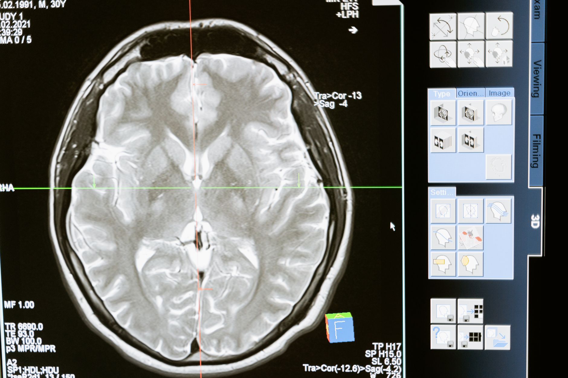
Researchers from Sweden have reported that women who experience a longer period of endogenous estrogen exposure, as well as those who use menopausal hormone therapy, exhibit fewer indicators of small‑vessel disease in the brain when they reach their early seventies. The findings, published in the journal Neurology, add to a growing body of evidence that hormonal fluctuations across a woman’s life may influence cerebrovascular health later in life.
Study design and participants
The analysis drew on data from the Gothenburg H70 Birth Cohort Studies, a longitudinal project that follows individuals born in 1944. A total of 352 healthy, cisgender Swedish women were included. Each participant underwent magnetic resonance imaging (MRI) at age 70, and most returned for a follow‑up scan at age 75. Researchers collected self‑reported information on the length of each woman’s reproductive years—calculated as the age at menopause or bilateral oophorectomy minus the age of menarche—as well as histories of oral contraceptive use and menopausal hormone therapy. These self‑reports served as proxies for lifetime estrogen exposure, though actual hormone levels were not measured.
Findings on vascular health
Analysis of the MRI scans focused on markers of small‑vessel disease, including white‑matter hyperintensities, lacunes, cerebral microbleeds, and enlarged perivascular spaces in the basal ganglia. The study found that each additional year of reproductive lifespan reduced a woman’s odds of presenting two or more such markers at age 70 by roughly 8 percent. The protective effect was driven primarily by later onset of menopause rather than an earlier age at first period.
Women who reported using menopausal hormone therapy also showed a lower burden of small‑vessel disease and better microstructural integrity of white matter. Notably, among participants carrying the APOE‑ε4 allele—a genetic variant that raises Alzheimer’s disease risk—hormone‑therapy users demonstrated greater cortical thickness in regions typically vulnerable to Alzheimer’s pathology. No comparable association was observed in women without the APOE‑ε4 variant.
Neurobiologist Anna Marseglia of the Karolinska Institutet, a senior author of the study, explained, “Our results suggest that hormonal exposure across different periods of a woman’s life may have cumulative effects also on the cerebrovascular health in later life.” She added that the brain’s blood vessels may be a key link between estrogen and brain aging.
Implications and limitations
Although the observational design precludes definitive statements about causality, the authors argue that longer endogenous estrogen exposure and a history of hormone therapy could support cerebrovascular integrity, potentially benefiting brain health in late life. The study’s authors caution that many associations lost statistical significance when stricter thresholds were applied, and they acknowledge that other hormonal changes during menopause may also play a role.
Yale neuroscientist Giulia Lorenzon, the study’s first author, emphasized that the results should not be interpreted as a recommendation for hormone therapy to prevent cognitive decline. She referenced a major review that found no strong evidence that menopausal hormone therapy alters dementia risk, either positively or negatively.
Given the limited size of the cohort and its focus on a single country, the researchers note that further work is needed to determine whether the observed patterns hold in more diverse populations. They conclude that clarifying estrogen’s long‑term impact on brain structure and cognition remains an important clinical priority, especially as personalized medicine seeks sex‑specific prevention strategies for cognitive disorders.

Mitchell Landsberg is a Senior Technology Correspondent at News Raise. He covers consumer electronics, artificial intelligence, software developments, and digital privacy trends.