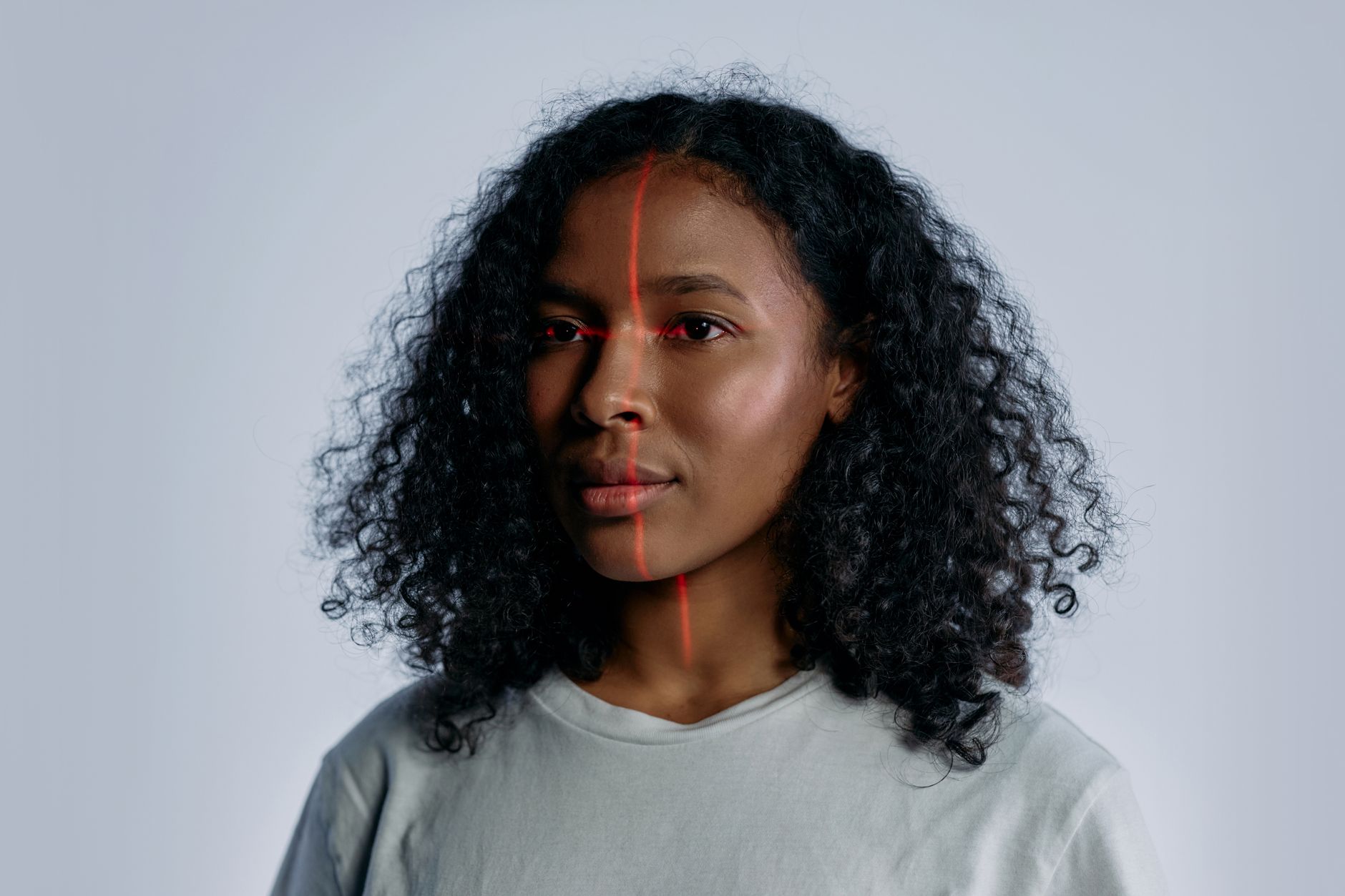
Scientists at Mass General Brigham in Boston have demonstrated that a single facial photograph can reveal clues about a cancer patient’s biological age and short‑term survival probability. The findings, published in The Lancet Digital Health, describe two artificial‑intelligence models that analyze facial features to estimate how a patient’s body has aged at the cellular level and to forecast mortality risk within 90 days.
How the models work
The research team first created a broad AI system called FAHR‑Face (Foundation Artificial Intelligence for Health Recognition‑Face), training it on more than 40 million images that represented a wide range of ages and ethnicities. From this foundation they derived two specialized models. FAHR‑FaceAge predicts a person’s biological age based on regions such as the temples, forehead, area above the eyes and the nasolabial fold that runs from the nose to the mouth. FAHR‑FaceSurvival, in contrast, focuses on the bridge of the nose and the area under the eyes to generate a mortality risk score that is then combined with standard clinical data.
According to study author Dr. Raymond Mak, a radiation oncologist at the Mass General Brigham Cancer Institute, the algorithm’s attention to these facial zones mirrors descriptions found in Hippocrates’ ancient “Book of Prognostics,” which linked a sharp nose, sunken eyes and fallen temples to declining health.
Performance on cancer patients
When applied to a cohort of cancer patients receiving radiotherapy, the FAHR‑FaceSurvival model identified 122 individuals as high‑risk; more than half of this group (52.5 %) died within 90 days of assessment. By comparison, only about 2 % of the 975 patients classified as low‑risk experienced death in the same period.
The biological‑age model showed a similar pattern. Among 1,060 patients whose estimated biological age exceeded their chronological age by more than 20 years, 21 % died within 90 days. In contrast, just 6 % of the 4,195 patients whose biological age was at least a decade younger than their actual age faced mortality in that window.
Researchers also reported that cosmetic alterations—such as facial surgery, injectable fillers or makeup—did not meaningfully impair the models’ predictive accuracy.
Clinical implications and expert commentary
The authors stress that the AI tools are intended to supplement, not replace, physician judgment. Dr. Mak suggests that early detection of accelerated aging or elevated mortality scores could prompt additional testing or earlier interventions for serious conditions.
External experts highlighted both promise and caution. Dr. Wilbur Lam, a professor at Emory University, called the study “extremely compelling” but noted that prospective validation is required before the technology can be trusted in everyday practice. He also raised questions about performance across diverse facial features, skin tones and age groups.
Professor Miroslaw Bober of the University of Surrey echoed the need for further testing, pointing out that while the AI can statistically link facial appearance to outcomes, it does not directly measure underlying biological aging processes. He warned that the predictive ability does not equate to explanatory insight and that the tool must be shown to add value beyond existing clinical assessments.
Overall, the research provides “convincing evidence” of a statistical relationship between facial cues and short‑term survival among radiotherapy patients, but the authors and commentators agree that broader, prospective studies are essential before integration into standard oncology care.

Steve Lopez is a Senior Editorial Columnist and Health & Public Policy reporter for News Raise. Steve focuses on healthcare advancements, medical technologies, and public health policies.